Magnetic Resonance - Technology Information Portal Welcome to MRI Technology
Info
  Sheets

Out-
      side


 'Active Shimming' 
SEARCH FOR    
 
  2 3 5 A B C D E F G H I J K L M N O P Q R S T U V W X Y Z
Result : Searchterm 'Active Shimming' found in 1 term [] and 3 definitions [], (+ 17 Boolean[] results
previous     6 - 10 (of 21)     next
Result Pages : [1]  [2 3 4 5]
MRI Resources 
MR Myelography - MRI Technician and Technologist Schools - Most Wanted - Spine MRI - Research Labs - Collections
 
Signa SP 0.5T™ Open ConfigurationInfoSheet: - Devices -
Intro, 
Types of Magnets, 
Overview, 
etc.MRI Resource Directory:
 - Devices -
 
www.gehealthcare.com/usen/mr/index.html From GE Healthcare;
The Signa SP 0.5T™ is an open MRI magnet that is designed for use in interventional radiology and intra-operative imaging. The vertical gap configuration increases patient positioning options, improves patient observation, and allows continuous access to the patient during imaging. The magnet enclosure also incorporates an intercom, patient observation video camera, laser patient alignment lights, and task lighting in the imaging volume.
Device Information and Specification
CLINICAL APPLICATION
Whole body
CONFIGURATION
Open
Integrated transmit and receive body coil; optional rotational body coil, head; other coils optional; open architecture makes system compatible with a wide selection of coilsarray
SYNCHRONIZATION
Standard cardiac gating, ECG/peripheral, respiratory gating
PULSE SEQUENCES
Standard: SE, IR, 2D/3D GRE and SPGR, 2D/3D TOF, 2D/3D FSE, 2D/3D FGRE and FSPGR, SSFP, FLAIR, EPI, optional: 2D/3D Fiesta, true chem sat, fat/water separation, single shot diffusion EPI
IMAGING MODES
Localizer, single slice, multislice, volume, fast, POMP, multi slab, cine, slice and frequency zip, extended dynamic range, tailored RF
TR
1.3 to 12000 msec in increments of 1 msec
TE
0.4 to 2000 msec in increments of 1 msec
3cm to 40 cm continuous
2D: 1.4mm - 20mm 3D: 0.2mm - 20mm
1280 x 1024
MEASURING MATRIX
128x512 steps 32 phase//freq.
PIXEL INTENSITY
256 gray levels
60 x 58 cm
POWER REQUIREMENTS
200 - 480, 3-phase
COOLING SYSTEM TYPE
Liquid helium
0.03 L/hr, holds 300 L
STRENGTH
12 mT/m
Active
spacer
 
Further Reading:
  News & More:
MR Surgical Suite, Improving surgical procedure quality (.pdf)
   by www3.gehealthcare.com    
MRI Resources 
Pediatric and Fetal MRI - MRA - Used and Refurbished MRI Equipment - Brain MRI - Services and Supplies - Case Studies
 
MAGNETOM Prisma
 
www.healthcare.siemens.com/magnetic-resonance-imaging/3t-mri-scanner/magnetom-prisma 
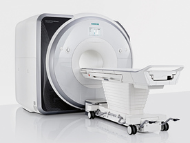
From Siemens Medical Systems; Received FDA clearance in 2013.
The MAGNETOM Prisma is the 3T PowerPack for exploration that offers most demanding clinical and research challenges of today and the future. The latest parallel transmit technology, TimTX TrueShape, enables zooming into specific body regions for enhanced image quality. Furthermore, the Tim 4G integrated coil technology offers remarkable imaging flexibility and supports complex examinations across the whole body.
Onsite upgrades to the MAGNETOM Prisma for customers who have already installed the 3 Tesla MAGNETOM Trio are possible.
Device Information and Specification
CLINICAL APPLICATION
Whole Body
CONFIGURATION
Ultra-short bore
3 Tesla
Head, spine, torso/ body coil, neurovascular, cardiac, neck, shoulder, knee, wrist, foot//ankle and multi-purpose flex coils. Peripheral vascular, breast, shoulder.
CHANNELS (min. / max. configuration)
64, 128
IMAGING TECHNIQUES
iPAT, mSENSE and GRAPPA (image, k-space), noncontrast angiography, radial motion compensation, Dixon
FOV
0.5 - 50 cm
BORE DIAMETER
or W x H
At isocenter: L-R 60 cm
TABLE CAPACITY
250 kg
MAGNET WEIGHT (gantry included)
13000 kg
DIMENSION H*W*D (gantry included)
173 x 230 x 222 cm
5-GAUSS FRINGE FIELD
2.6 m / 4.6 m
CRYOGEN USE
Zero boil off rate, refill approx. 10 years
COOLING SYSTEM
Water
up to 200 T/m/s
MAX. AMPLITUDE
80 mT/m
Passive, active; first order, second order
POWER REQUIREMENTS
380 / 400 / 420 / 440 / 460 / 480 V, 3-phase + ground;
spacer
MRI Resources 
Education pool - Fluorescence - Databases - Pacemaker - MRI Accidents - Breast Implant
 
Excelart AG™ with PianissimoInfoSheet: - Devices -
Intro, 
Types of Magnets, 
Overview, 
etc.MRI Resource Directory:
 - Devices -
 
http://www.medical.toshiba.com/clinical/radiology/15texcelart.htm From Toshiba America Medical Systems Inc.;
the EXCELART is a superconducting whole body MRI system with a short wide-bore magnet, operating at 1.5 T. It features powerful high-speed gradients with a revolutionary gradient acoustic noise reduction system: Pianissimo. The dramatic reduction of gradient acoustic noise by Pianissimo greatly enhances patient comfort during exams. The standard array platform and a wide range of array coils ensure excellent images. A powerful 64-bit RISC-based computer system and newly developed array processor realize high productivity.
Device Information and Specification
CLINICAL APPLICATION
Whole body
CONFIGURATION
Cylindrical Wide Short Bore
Opt. (WIP) Single and Multi Voxel
SYNCHRONIZATION
ECG: standard; peripheral: optional, respiratory gating
PULSE SEQUENCES
SE, FE, IR, FastSE, FastIR, FastFLAIR, Fast STIR, FastFE, FASE, Hybrid EPI, Multi Shot EPI; Angiography: 2D(gate/non-gate)/3D TOF, SORS-STC
IMAGING MODES
Single, multislice, volume study
TR
3.5-30000 msec
TE
8 msec min. SE; 1.2 msec min. FE
SINGLE/MULTI SLICE
less than 0.015 (256x256)
FOV
2 cm
1.0 min. 2-DFT: 0.2 min. 3-DFT
Up to 1024
MEASURING MATRIX
32-1024, phase;; 64-1024, freq.
PIXEL INTENSITY
256 gray levels
BORE DIAMETER
or W x H
65.5 cm, patient aperture
MAGNET WEIGHT
4050 kg (bare magnet incl. L-He)
H*W*D
235 x 219 x 199 cm
COOLING SYSTEM TYPE
Closed-loop water-cooled
CRYOGEN USE
Liquid helium: approx. less than 0.05 L/hr
STRENGTH
25 mT/m
5-GAUSS FRINGE FIELD
2.5 m / 4.0 m
Passive, active, auto-active
spacer

• View the DATABASE results for 'Excelart AG™ with Pianissimo' (2).Open this link in a new window

MRI Resources 
Contrast Enhanced MRI - RIS - Education - General - Corporations - Societies
 
Echelon™ 1.5TInfoSheet: - Devices -
Intro, 
Types of Magnets, 
Overview, 
etc.
 
www.hitachimed.com/contentindex.asp?ID=971 From Hitachi Medical Systems America Inc.;
Hitachi expanded its portfolio with the Echelon™ 1.5T. The MRI scanner combines a compact magnet and a scalable 8-channel RF system with high-performance gradients and slew rate to select short echo times, small field of views, high matrices and thin slices. Standard features of the Echelon MRI system include higher-order active shim, RAPID (parallel imaging for use on brain MRI, body, cardiovascular imaging, and orthopedic coils), multiple coil ports, and an advanced reconstruction engine.
Device Information and Specification
CLINICAL APPLICATION
Whole body
CONFIGURATION
Short bore
Head, body coil, spine, breast, knee, shoulder, vascular multiple array coils.
SYNCHRONIZATION
Cardiac gating, ECG/peripheral, respiratory gating
PULSE SEQUENCES
SE, IR, FSE, FIR, GE, SG, BASG, PBSG, PCIR, DWI, Radial, Angiography: TOF, FLUTE (Fluoro-triggered bolus MRA), Time-resolved MRA
IMAGING MODES
Single, multislice, volume study
PIXEL INTENSITY
Level Range: -2,000 to +4,000
Sub millimeter
POWER REQUIREMENTS
208/220/240 V, single phase
CRYOGEN USE
Low cryogen boil-off
STRENGTH
30 mT/m
150 T/m/sec
Higher-order active shim
spacer

• View the DATABASE results for 'Echelon™ 1.5T' (2).Open this link in a new window


• View the NEWS results for 'Echelon™ 1.5T' (3).Open this link in a new window.
 
Further Reading:
  Basics:
Echelon 1.5T
   by www.hitachimed.com    
MRI Resources 
MRI Physics - Journals - Colonography - Process Analysis - MR Guided Interventions - Mobile MRI
 
Excelart XG™ with PianissimoInfoSheet: - Devices -
Intro, 
Types of Magnets, 
Overview, 
etc.MRI Resource Directory:
 - Devices -
 
http://www.medical.toshiba.com/clinical/radiology/15texcelart.htm From Toshiba America Medical Systems Inc.;
the EXCELART is a superconducting whole body MRI system with a short wide-bore magnet, operating at 1.5 T. It features powerful high-speed gradients with a revolutionary gradient acoustic noise reduction system: Pianissimo. The dramatic reduction of gradient acoustic noise by Pianissimo greatly enhances patient comfort during exams. The standard array platform and a wide range of array coils ensure excellent images. A powerful 64-bit RISC-based computer system and newly developed array processor realize high productivity.
Device Information and Specification
CLINICAL APPLICATION
Whole body
CONFIGURATION
Cylindrical Wide Short Bore
Optional (WIP)
SYNCHRONIZATION
ECG/peripheral, respiratory gating
PULSE SEQUENCES
SE, FE, IR, FastSE, FastIR, FastFLAIR, Fast STIR, FastFE, FASE, Hybrid EPI, Multi Shot EPI; Angiography: 2D(gate/non-gate)/3D TOF, SORS-STC
IMAGING MODES
Single, multislice, volume study
TR
2.6-30000 msec
TE
8 msec min. SE; 0.9 msec min. FE
SINGLE/MULTI SLICE
less than 0.011 (256x256)
FOV
2 cm
1.0 min. 2-DFT: 0.2 min. 3-DFT
Up to 1024
MEASURING MATRIX
32-1024, phase;; 64-1024, freq.
PIXEL INTENSITY
256 gray levels
BORE DIAMETER
or W x H
65.5 cm, patient aperture
MAGNET WEIGHT
4050 kg (bare magnet incl. L-He)
H*W*D
235 x 219 x 199 cm
POWER REQUIREMENTS
380/400/415/440/480 V
COOLING SYSTEM TYPE
Closed-loop water-cooled
CRYOGEN USE
Liquid helium: approx. less than 0.05 L/hr
STRENGTH
25 mT/m
5-GAUSS FRINGE FIELD
2.5 m / 4.0 m
Passive, active, auto-active
spacer

• View the DATABASE results for 'Excelart XG™ with Pianissimo' (2).Open this link in a new window

 
Further Reading:
  News & More:
Angio, cardiac imaging top list for MR and CT
1999   by www.diagnosticimaging.com    
MRI Resources 
Raman Spectroscopy - Health - Breast Implant - MR Guided Interventions - Calculation - Mobile MRI Rental
 
previous      6 - 10 (of 21)     next
Result Pages : [1]  [2 3 4 5]
 Random Page
 
Share This Page
FacebookTwitterLinkedIn

MR-TIP    
Community   
User
Pass
Forgot your UserID/Password ?    


Personalized protocols (age, gender, body habitus, etc.) lead to :
more automated planning 
improved patient comfort 
better diagnostics 
optimized image quality 
nothing 

Look
      Ups


MR-TIP.com uses cookies! By browsing MR-TIP.com, you agree to our use of cookies.

Magnetic Resonance - Technology Information Portal
Member of SoftWays' Medical Imaging Group - MR-TIP • Radiology-TIP • Medical-Ultrasound-Imaging • 
Copyright © 2003 - 2024 SoftWays. All rights reserved. [ 28 April 2024]
Terms of Use | Privacy Policy | Advertising
 [last update: 2024-02-26 03:41:00]